Mini-Course #9 | A neuroanatomy and neuro-function approach to the emergence and nature of consciousness during development. | Bernard Baars & Nat Geld
Consciousness may seem simple and obvious to many of us. When we’re awake, we are aware of the outer world and of our own inner thoughts and feelings. When we fall asleep, that inner and outer awareness slips away.
Our own consciousness is hard to study because we cannot vary it; as soon as we lose consciousness, we can no longer observe anything. And how can we observe someone else’s conscious experience? In Mini-Course #9, Dr. Jay Giedd and neuroscientist David Edelman talk candidly about our understanding of the complexities of the developing brain and consciousness.
David Edelman is a paleoanthropologist and neuroscientist focused on consciousness in non-human animals, and Dr. Jay Giedd is a practicing psychiatrist and neuroscientist at UCSD School of Medicine who has spent more than thirty years exploring the growth and development of the human brain, from embryogenesis through childhood and adolescence well into adulthood, in his groundbreaking National Institute of Mental Health (NIMH) study.
So this lesson explores a neuroanatomy and neuro-function approach to some fundamental issues that confront the science of consciousness. We consider the transition from wakefulness to deep non-REM sleep and its signal importance as a transition between conscious and non-conscious states during which changes in brain activity occur that we can actually study—and that provides clues as to the nature of consciousness.
How do we define consciousness? What does that term mean? Where do we even start? As a developmental neuropsychiatrist at UCSD School of Medicine and Rady Children’s Hospital, Dr. Jay Giedd wants to try to understand consciousness from a neuroanatomy and neuro-function standpoint, and asks:
“What would consciousness look like in a brain scanner and other types of imaging?
What are we looking for, in a sense, and could I predict from basically the architecture and the anatomy, that this could be conscious, and this would not be able to be conscious?"
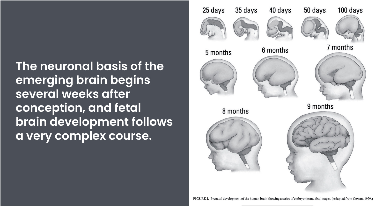
The neuronal basis of the emerging brain begins several weeks after conception, and fetal brain development follows a very complex course.1 How can we study it?
Treat consciousness as a variable.
Consciousness may seem simple and obvious to many of us. When we’re awake, we are aware of the outer world and of our own inner thoughts and feelings. When we fall asleep, that inner and outer awareness slips away.
We can only study something if we can treat it as a variable, comparing its presence to its absence.
A number of historic breakthroughs in science emerged from the realization that some previously assumed constant, like atmospheric pressure or gravity, was actually a variable. It required a great leap of imagination for natural philosophers to understand that all objects in the universe need not fall toward the earth, that gravity could be different elsewhere in the universe. Newton's ability to make that leap led to the solution of the ancient puzzle of planetary motion.
Consciousness has seemed to be different from all other scientific concepts; it has been extraordinarily difficult to treat it as a variable. The persistent pattern over centuries has been to see our own experience as the only psychological domain that can be conceived, one that has no kinship to any conceivable comparison condition.
Our own consciousness is hard to study because we cannot vary it; as soon as we lose consciousness, we can no longer observe anything. And how can we observe someone else's conscious experience?
It is thought that there must be a time when the emerging brain changes from a small, non-conscious collection of neurons to a massive, 86 billion-cell structure enabling conscious feelings, sensory perception and thoughts. When is that time of transition? Early or late in fetal development?
These are key questions that David Edelman and brain development specialist Jay Giedd examine as they discuss the emergence of consciousness in early life. Dr. Jay Giedd has spent more than thirty years studying the brain trajectory from embryo to fetus, then infancy and childhood, and then adolescence and adulthood.
Jay Giedd & David Edelman on the emergence and nature of consciousness during development.
Giedd and Edelman explore the idea that memory may be an essential aspect of conscious states. Very young infants experience the world with their developing senses, remembering some things and allowing memory traces to shape later experiences.
Changes in observable brain activity provide clues as to the nature of consciousness. Memories are not just images of the past—at the level of neurons, memories are connections between cells, and conscious experiences change the brain.
When do the first useful memories actually form?
There is certainly a Rubicon that is crossed; we just haven’t figured out when it happens or what that passage looks like. Memory is a ubiquitous faculty across the animal kingdom; even relatively simple animals like the humble marine snail Aplysia* can learn and remember at a fundamental level.
1) Aplysia Californica emitting an ink cloud. 2) Aplysia neurons (A,B) and growth cones (C) are some of the largest in the animal kingdom.
Aplysia Californica emitting an ink cloud. Aplysia dactylomela is one of the most abundant and cosmopolitan species of sea slugs. Charles Darwin, in January 1831, made a vivid description of this species at his very first stop on St. Jago (Cape Verde Islands) during the Voyage of the Beagle. Aplysia neurons (A,B) and growth cones (C) are some of the largest in the animal kingdom.2
*What is Aplysia? A genus of gastropod molluscs well-known as ‘model organisms’ in neuroscience, particularly work on the cellular biology of learning and memory (for his contributions Eric Kandel shared the 2000 Nobel prize for Physiology or Medicine). Their latin name comes from L’Aplysia — “that which one cannot wash”.
Aplysia (Figure 1) has one of the earliest mentions in the literature of any animal, the first authentic description being by Pliny in his Historia Naturalis, c. 60 A.D. They are also commonly called sea hares because their posterior chemosensory tentacles, the rhinophores, stick up like ears.
Is early human memory comparable to the kinds of memory we see in other animals like Aplysia?
The scalp EEG measures electromagnetic signals from the brain, and it has been known for many years that waking consciousness shows low voltage, fast, and irregular waveforms. At the opposite pole of deep sleep, called Slow-Wave Sleep, the scalp EEG shows high voltage, slow, and more regular peaks and valleys.
Because the human scalp blocks most of the brain signals (like 99.9%), it is only recently that scientists have started to understand the details of connectivity and traffic flow in Slow-Wave Sleep and conscious waking.
Scalp EEG is performed by placing small metal electrodes directly on the scalp, and measuring the electrical voltage that is picked up.
Neurons carry an electrical charge and they communicate by sending electrical and chemical signals to other neurons.
But the human scalp is very thick and it is covered by skin and blood vessels and layers of tissue and this barrier blocks most of the electrical activity from the brain.
The scalp EEG of dreaming states has been called “paradoxical” because they show small, irregular voltages, like conscious waking, but we are not conscious of external events. Rather, in dreams, our experiences are generated by the brain itself, as if the brain is talking to itself.
Sigmund Freud believed that the interpretation of dreams are “the royal road to the unconscious,” which has been interpreted to mean that unconscious goals and emotional processes may be expressed in dream experiences. We usually remember only a small fraction of our dreams.
Consciousness and learning
We will build on these basic ideas in Mini-Course #9, and explore:
Consciousness in the early growing brain.
When in life development are retrievable memories established?
The electrical activity of the brain (EEG) is a good indicator of mental processes.
But scalp EEG shows a 99.9 percent signal loss, so that more accurate measures are much more revealing. The surface EEG still suggests that “consciousness is a difference that makes an observable difference,” even at a fairly gross level. Consciousness is not some otherworldly halo that is separate from our mental experiences or their biological basis in the brain.
Is memory needed for the development of conscious functions?
Sleep is essential for useful memory formation.
Brain activity looks very different in sleep and waking.
The role of dreams — some knowns and unknowns.
🧩 The Developing Brain & Consciousness Quiz
This short quiz will help you develop some of the key ideas from Mini-Course #9. Lean in and enjoy — learning is fun!
Question 1
Which of the following are similar?
A) Waking and sleeping EEG
C) Slow wave sleep (SWS) and dreaming sleep
B) EEG and EKG
D) Waking and REM dreams
✅ Answer: D
Question 2
How much of the raw brain signal is blocked during an EEG?
A) About half
B) About two-thirds
C) 99.9 percent
D) Not very much
✅ Answer: CScalp EEG is performed by placing small metal electrodes directly on the scalp, and measuring the electrical voltage that is picked up. Neurons carry an electrical charge and they communicate by sending electrical and chemical signals to other neurons. But the human scalp is very thick and it is covered by skin and blood vessels and layers of tissue. This barrier blocks most of the electrical activity from the brain.
Question 3
Because sleep makes us vulnerable to predators, how do various animals cope with that danger?
A) Dolphins actually sleep with one side of the brain at a time, so that they can keep swimming and maybe respond to danger.
B) Many animals do not have the deepest kind of sleep, Slow Wave Sleep.
C) Cats take catnaps.
D) Many animals show waking EEG throughout the night.
✅ Answer: A If like us, dolphins went into a deep unconscious sleep, they would stop breathing and suffocate or drown. To get around this, dolphins only allow one half of their brains to sleep at a time; the other half stays alert to enable the dolphin to continue breathing and look out for dangers in the environment.
Namaste. In a very long tradition, we salute the conscious self in you from our own conscious selves. We hope you enjoy exploring the conscious brain with us! :*) bjb 🧠✨
Giedd JN, Blumenthal J, Jeffries NO, Castellanos FX, Liu H, Zijdenbos A, Paus T, Evans AC, Rapoport JL. Brain development during childhood and adolescence: a longitudinal MRI study. Nat Neurosci. 1999 Oct;2(10):861-3. doi: 10.1038/13158. PMID: 10491603.
Lenroot RK, Giedd JN. Brain development in children and adolescents: insights from anatomical magnetic resonance imaging. Neurosci Biobehav Rev. 2006;30(6):718-29. doi: 10.1016/j.neubiorev.2006.06.001. Epub 2006 Aug 2. PMID: 16887188.
Figure 1 and 2: Photographs by Leonid L. Moroz (A,C) and Sami Jezzini (B).
Figure 2: (A) A photograph of the freshly dissected right abdominal semi-ganglion from A. californica with the positions of two neurons marked (R2 and R14). This R2 cell is the largest neuron ever photographed reaching 1.1 mm in diameter. When this cell was isolated, yielding a record 1.965 μg of total RNA (while typical mammalian neurons contain less than 50 pg of RNA). These cells can also provide from 150 to 250 ng of DNA depending upon cell size. (B) R2 neurons can be seen by the naked eye on a penny. (C) Giant growth cone of a single motoneuron in cell culture (2nd day) stained with anti-β-tubulin antisera (red). Photographs by Leonid L. Moroz (A,C) and Sami Jezzini (B).